
Loss of control or leakage
Shows muscle shape and continuity to relate continence anatomy to symptoms.
Imaging the muscles of bowel control to plan your treatment
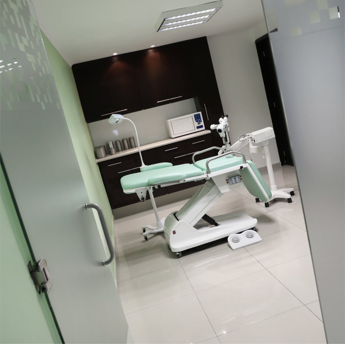
360° endoanal ultrasound shows the sphincter and surrounding tissue. We offer it in the office, avoiding travel elsewhere and returning with results, for greater convenience and continuity of care.
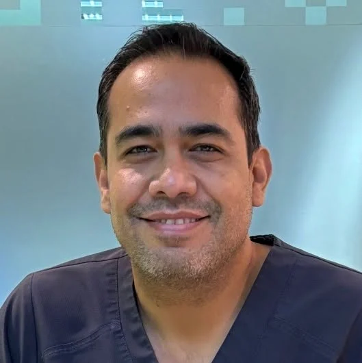
Dr. Eduardo Navarro LaraColorectal surgeon · Anorectal tests
What it shows
The test takes place in the office, without needing to go elsewhere and return with results.
An ultrasound probe in the anal canal creates cross-sectional images. The 360° view examines the full circumference and relationships between internal and external sphincters and nearby tissue.
The clinical concern and examination determine how this anatomical information is used.
When it adds information
Symptoms, examination and history determine whether imaging adds useful information.
Shows muscle shape and continuity to relate continence anatomy to symptoms.
Identifies internal or external sphincter changes after childbirth, surgery or injury.
In selected cases, it follows fistula tracts and their muscle relationship before treatment decisions.
Images nearby collections and tissues when examination needs supplementation.
Tests answering different questions
No single test covers all colon, rectal and anal problems.
Shows sphincter anatomy and structures near the anal canal.
Directly views anal canal and lower rectal lining.
Explore anoscopyProvides a broader map for complex fistulas or extensions distant from the canal.
Colonoscopy serves another purpose: viewing the colon's interior. Learn about colonoscopy
From assessment to result
Indication and interpretation matter as much as the image, avoiding testing by name alone.
Check availabilityThe doctor relates symptoms, examination and prior tests to whether imaging answers the question.
The team confirms preparation, location and personal considerations before scheduling.
An ultrasound probe shows sphincters and nearby tissues in a 360° view.
Images are interpreted with the clinical concern to choose treatment, follow-up or further assessment.
Possible findings
An isolated finding is not the whole story. Its value comes from symptoms, function and history.
Imaging shows muscle rings contributing to anal closure.
Locates discontinuities or anatomical changes related to childbirth, injuries or previous surgery.
In selected cases, identifies the course and sphincter relationship.
Complements inflammation or abscess assessment without replacing active infection care.
Before the test
The team adapts the protocol for pain, inflammation, previous surgery or history and confirms preparation and location before scheduling.
Clinical interpretation
Assessment integrates imaging, examination, history and other tests to define treatment or further investigation.
Specialist care
Dr. Eduardo Navarro Lara is a colorectal surgeon offering 360° endoanal ultrasound in his current practice.
Assessment determines whether the test addresses your problem and interprets results within treatment options.
Explore his training and experienceCare for patients in Guadalajara, Zapopan and the metropolitan area
Our support team checks appointments at Unidad Médica Shakespeare or Torre de Consultorios Hospital Ángeles Andares. Test location and scheduling are confirmed during care.
Frequently asked questions
The probe images the full anal canal circumference, showing cross-sectional relationships between sphincters and surrounding tissues.
It checks sphincter injury or anatomical changes, interpreted alongside symptoms and functional tests.
Yes. In selected cases it shows the tract and sphincter relationship. Complex fistulas may need MRI or further assessment.
No. Anoscopy views the lining directly; ultrasound images surrounding muscles and tissues.
No. Colonoscopy views inside the colon; endoanal ultrasound focuses on sphincters and tissues around the anal canal.
Probe insertion may cause discomfort. Discuss severe pain, inflammation or active infection beforehand for suitable instructions.
The team adapts preparation to its protocol and your situation. Do not use enemas or stop medication from general advice.
Simply ask about appointment times and availability. Clinical details and previous tests are reviewed through the appropriate channel.
360° endoanal ultrasound
Request assessment with Dr. Eduardo Navarro to review the test's purpose and confirm availability.